 CASE REPORT
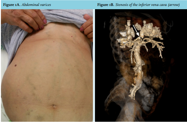
A 64-year-old female presented at our center with diffuse abdominal pain. Since her second decade of life, her medical history has been significant for abdominal (figure 1A) and leg venous varicosities with spontaneous recurrent skin bleeding episodes. Endoscopy showed esophageal varices. Laboratory values revealed a low platelet count of 73,000 x 103 /l, but were otherwise unremarkable with normal albumin level and (active) partial thromboplastin time serology results for hepatitis A, B, and C were negative. Abdominal ultrasound demonstrated normal liver size and texture, without evidence of ascites. Splenomegaly was found measuring 13.6 cm. Computed tomography (CT) angiography revealed a narrowing of the suprahepatic portion of the inferior vena cava (IVC) (figure 1B) with venous collaterals, but no evidence of thrombosis. Percutaneous balloon angioplasty with stent placement to the stenotic suprahepatic inferior vena cava was performed. The one-year follow-up visit revealed an increase in platelet count to 100,000 x 103 /l, a decrease in spleen size to 10.5 cm, no episodes of leg varicosity bleeding, as well as an improvement in the esophageal varices grade.
WHAT IS YOUR DIAGNOSIS?
See page 269 for the answer to this photo quiz.