Full textPDF
Full text
CASE REPORT 
A 44-year-old previously healthy woman was admitted to the hospital with complaints of acute abdominal pain, anorexia, and sweating, which started one week prior to admission. An abdominal computerized tomography (CT) scan revealed a mass in the left ovary (10 x 14 cm), a second mass in the left adrenal gland (5 x 6 cm) and massive ascites throughout the abdomen. Laboratory investigations revealed a CA 125 level of 706 U/l (normal < 35 U/l), lactate dehydrogenase (LDH) of 1279 U/l (normal < 250 U/l), and normal haemoglobin, platelets, leukocytes, kidney function, and liver enzymes. Cytology of the ascites showed macrophages loaded with haemosiderin and a few mesothelial cells, lymphocytes and blood cells, but no malignant cells.
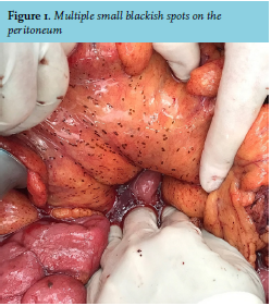
Upon laparotomy, there were 3 litres of dark ascites. Multiple, small, non-palpable blackish spots were visible on the peritoneum (figure 1). The left ovary was enlarged (10 x 20 cm) and dark black. We performed a hysterectomy and bilateral salpingectomy, and an omentectomy and biopsies of peritoneum and adrenal gland.
WHAT IS YOUR DIAGNOSIS?
See page 125 for the answer to this photoquiz.

