Full textPDF
Full text
CASE REPORT 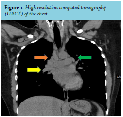
A 33-year-old female of south Indian origin presented with a history of low-grade fever, migratory polyarthralgia with redness and swelling in her legs. On examination, there were erythematous, tender, immobile nodules over both of her shins. The remainder of her general and systemic examination were unremarkable. Her investigation revealed elevated serum angiotensin converting enzyme (ACE) level along with a non-reactive Tuberculin skin test and negative quantiFERON-TB Gold (QFT). High resolution computed tomography (HRCT) of the chest revealed a combination of right paratracheal nodes (figure 1, orange arrow), right hilar nodes (figure 1, yellow arrow) and left hilar nodes (figure 1, green arrow). Identify this sign, which is very typical of a particular condition and described by chest X-ray.
WHAT IS THE DIAGNOSIS?
See page 35 for the answer to this photo quiz.