Full textPDF
Full text
CASE REPORT 
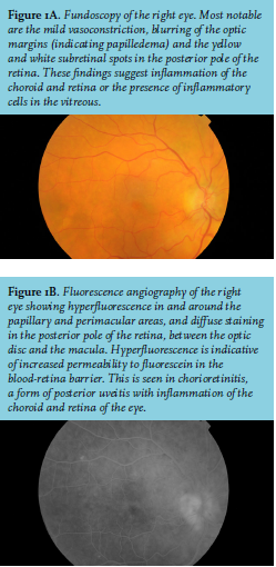
A 74-year-old male was admitted to the hospital complaining of progressive visual decline. Medical history revealed diabetes mellitus, atrial fibrillation, hypercholesterolemia and gout. He worked as a crane operator in the harbor of Rotterdam, the Netherlands. At presentation, he reported a weight loss of 10 kg in the last three months. Visual acuity was 0.16 in both eyes. Initial laboratory investigation revealed a significantly raised erythrocyte sedimentation rate (128 mm/h) but otherwise no abnormalities. Computed tomography and magnetic resonance imaging of the brain showed no significant abnormalities. The ophthalmologist performed fundoscopy and fluorescein angiography (figures 1A and 1B).
WHAT IS YOUR DIAGNOSIS?
See page 453 for the answer to this photo quiz

