Full textPDF
Full text
CASE REPORT
A 50-year-old male presented to our outpatient clinic after referral for a cardiac catheterisation for a three-month history of stable angina pectoris. He described his chest pain as dull, oppressive in quality and related to physical effort. His medical history was remarkable for diabetes mellitus, hypertension, and high blood cholesterol. He had also undergone thoracic surgery at the age of 12, but was unable to remember more details about it.
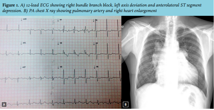
Cardiac examination demonstrated a laterally displaced point of maximum impulse, a left lower parasternal heave and a grade IV pulmonary systolic-diastolic murmur. ECG revealed right bundle branch block, left axis deviation, slight ST-segment elevation in aVR, and negative T waves and ST-segment depression in V3-V6 and I-aVL (figure 1A). A chest X-ray showed pulmonary artery and right heart enlargement (figure 1B).
WHAT IS YOUR DIAGNOSIS?