DIAGNOSIS
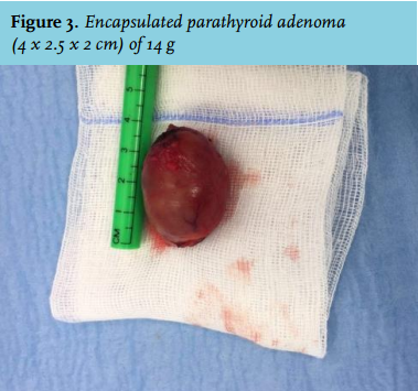
The multifocality of the bone lesions raised the suspicion of malignancy. Therefore, the patient was referred for biopsy of the lesion of the left third rib. Microscopy revealed numerous multinucleated giant cells compatible with a brown tumour of the bone. No signs of malignancy were revealed. Minimal invasive parathyroidectomy was performed disclosing a large parathyroid gland of 14 g (4.0 x 2.5 x 2.0 cm) (figure 3). The gland was perfectly encapsulated. Histology revealed a follicular and trabecular architecture of chief cells. Between these cells, multiple vascular structures and fibroblasts were present. Postoperatively, the calcium and parathyroid hormone (PTH) levels rapidly normalised.
This is one of the first demonstrations of multiple FDG-avid lesions of brown tumours due to hyperparathyroidism.1 The histological features of brown tumours consist of mononuclear stromal cells as well as multinucleated giant cells. The brown colour originates from haemorrhagic infiltrates and haemosiderin deposits by macrophages in response to bone destruction. The bone defect fills with fibroblastic tissue that can deform the bone structure and may resemble a neoplastic process.2 The elevated FDG uptake in this process can be explained by the high bone turnover, immunogenic activity and inflammatory response by haematopoietic cells, fused osteoclasts (multinuclear giant cells) and macrophages.
Skeletal changes due to high PTH exposure result in bone pain and arthralgia along with high hypercalcaemia as in our case.3 PET/CT imaging is a novel technique for the discrimination of FDG-avid from non-avid lesions but histology remains the cornerstone for the final diagnosis of either brown tumour lesions or bone metastases.
CONCLUSION
FDG-avid lesions of brown tumours can mimic skeletal metastases in patients with hyperparathyroidism. The diagnosis of brown tumours should always be considered in patients with primary hyperparathyroidism and multiple bone lesions.
REFERENCES