Full textPDF
Full text
CASE REPORT
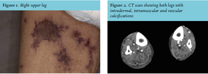
A 66-year-old male haemodialysis patient with chronic kidney failure caused by hypertension presented at the emergency department after being found on the floor at home. He had multiple comorbidities and was known to be noncompliant to his prescribed medication. He suffered from pain in both upper legs and was not able to get up by himself. At admission his international normalised ratio was 1.2, potassium 5.5 mmol/l, phosphate 4.1 mmol/l, calcium 2.59 mmol/l and parathyroid hormone 137.0 pmol/l. He was taking acenocoumarol for atrial flutter. We noticed a colour change at multiple sites on his upper legs, which later progressed into blue discolorations with necrosis and palpable, very painful skin lesions several centimetres wide in arbitrary areas (figure 1). Duplex ultrasonography and computer tomography of the abdominal and large arteries of the limbs revealed vascular wall calcification in all the major vessels but no vascular occlusions (figure 2).
WHAT IS YOUR DIAGNOSIS?