Full textPDF
Full text
CASE REPORT
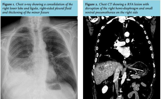
A 76-year-old man was referred to the hospital with progressive dyspnoea and abdominal pain in the right upper quadrant of the abdomen. Medical history showed colon carcinoma with metastasis of the liver, for which radiofrequency ablation had been performed several times, most recently three weeks prior to presentation. Physical examination showed a temperature of 38.6 °C and respiratory rate of 32 breaths/min with diminished breath sounds at the right basal area with fine rales and abdominal tenderness of the right upper quadrant. Blood analysis showed a C-reactive protein level of 92 mg/l (normal value 0-10mg/l) and a leukocyte count of 14.2 x 10⁹/l (normal value 3.5-10.0 x 10⁹/l). Chest X-ray showed consolidation of the right lower lobe and ligula, right-sided pleural fluid and thickening of the minor fissure (figure 1). Computed tomography, performed after drainage of the pleural fluid, showed a lesion in the liver with air and a perforation of the right hemi-diaphragm with a small ventral pneumothorax (figure 2).
WHAT IS YOUR DIAGNOSIS?