Full textPDF
Full text
 CASE REPORT
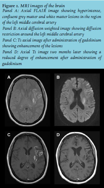
A 49-year-old woman presented to the Emergency Department with aphasia; the time of onset was unknown. Her past medical history revealed a syphilis infection in 1987 and alcohol abuse. On neurological examination she spoke mainly non-existent words, her comprehension was relatively unaffected. Furthermore, there was a mild right-sided central facial palsy, right-sided hyperreflexia, and bilateral Babinski signs. Computed tomographic imaging of the brain showed a hypodense lesion in the left hemisphere. Magnetic resonance imaging of the brain revealed hyperintense, confluent grey matter and white matter lesions in the left hemisphere (figure 1). The lesions were isointense on T1 weighted images and enhanced after administration of gadolinium.
WHAT IS YOUR DIAGNOSIS?

