Full textPDF
Full text
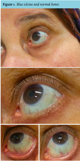 CASE
CASE
A 64-year-old Moroccan woman was referred to our outpatient clinic because of extreme fatigue. Her medical history revealed hypertension and femoral deep venous thrombosis (DVT). Furthermore, she had been using oral iron supplementation for anaemia due to iron deficiency for seven years. She suffered from occasional gastro-oesophageal reflux disease. Her stools were normal, without macroscopic blood, but black-coloured. She had no vaginal blood loss. Despite the fact that the DVT occurred two years earlier, she was still using oral anticoagulation. Her family history was unremarkable. Physical examination showed distinct blue sclerae with moderately pale conjunctivae. Abdominal and rectal examination revealed no abnormalities. Laboratory results demonstrated haemoglobin 4.8 mmol/l, mean cell volume 65 fl, ferritin 7 µg/l, transferrin saturation 3%, reticulocytes 40.8 x 10⁹/l, normal levels of thrombocytes and leukocytes, haptoglobin, lactate dehydrogenase, vitamin B12 and folic acid. Renal and thyroid function was normal.
WHAT IS YOUR DIAGNOSIS?