KEY WORDS 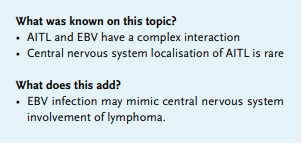
Angioimmunoblastic T-cell lymphoma (AITL), central nervous system involvement, Epstein-Barr virus (EBV), mimicking lymphoma
INTRODUCTION
The clinical picture of Epstein-Barr virus (EBV) infection may vary from largely asymptomatic patients to a dramatic clinical course, which may include EBV-related meningoencephalitis. EBV infection and malignant lymphoma share a variety of clinical and pathological features, which may result in an incorrect diagnosis of malignant lymphoma.1-4
We present a patient with an unusual course of EBV infection, who was initially diagnosed with angioimmunoblastic T-cell lymphoma (AITL) with central nervous system (CNS) localisation. This case demonstrates the complexity of the clinical picture and the subsequent use of additional investigations leading to the correct diagnosis.
CASE REPORT
A 51-year-old man without a significant medical history presented with headache, episodic confusion, nausea, vomiting, fatigue, fever, night sweats, and weight loss following a minor head injury. Physical and neurological examination was normal. A computed tomography (CT) scan of the head was normal. The cerebrospinal fluid (CSF) showed 112 x 106/l leukocytes (100% lymphocytes) and an elevated total protein of 1.7 g/l, suggestive of a viral meningoencephalitis, which was treated empirically with intravenous acyclovir. Virological analysis of the CSF showed an EBV deoxyribonucleic acid (DNA) load of 23605 IU/ml, while EBV DNA in serum was 30-fold lower (794 IU/ml). Flow cytometry of the CSF identified primarily T lymphocytes, without clear monoclonality. Serum lactate dehydrogenase was 494 U/l, whereas alanine aminotransaminase and aspartate aminotransaminase were also elevated (194 and 140 U/l, respectively). A positron emission tomography (PET) scan showed diffuse fluor-18-deoxy glucose (FDG) avid lymphadenopathy (cervical, axillar, mediastinal, retroperitoneal, para-iliacal, and inguinal) with splenomegaly. Biopsies of both tonsil and axillary lymph node yielded a pathological preferential/working diagnosis of angioimmunoblastic T-cell lymphoma (AITL) with expression of pan T-cell antigens (CD3, CD4, CD5) and positive EBV-encoded small ribonucleic acids. Pending the results of gene rearrangement studies, the clinical working diagnosis was AITL with CNS localisation and the patient was transferred to a tertiary referral centre for further workup and treatment. Further evaluation with a magnetic resonance imaging scan showed no intracranial abnormalities. In the bone marrow, a reactive polyclonal T-cell population was observed. Repeated CSF analyses revealed a leucocyte count of 17 x 106/l (100% lymphocytes), a total protein of 0.61 g/l, and normal flow cytometry. Strikingly, EBV DNA was negative in both CSF and serum. Upon revision of the lymph node histology, the results of the gene rearrangement analysis demonstrated a polyclonal B-cell and T-cell population. Hence, a watchful waiting approach was adopted. Clinically, the patient completely recovered. In addition, follow-up FDG-PET scan showed quasi-normalisation except for some residual FDG activity in the axillary lymph nodes, confirming the definitive diagnosis of a systemic EBV infection with secondary EBV meningoencephalitis.
DISCUSSION
AITL is a rare and aggressive subtype of a peripheral T-cell lymphoma, classified as a nodal T-cell lymphoma with T-follicular helper phenotype, according to the 2016 World Health Organisation classification.5 It accounts for approximately 1-2% of non-Hodgkin’s lymphoma.6,7 Central nervous system involvement is rarely reported.8 AITL has been known to have a complex relationship to EBV.7,9,10 The initiating event of lymphoma development has been suggested to be antigen driven by EBV because EBV-positive B cells are found early in the course of the disease.11 In addition, it has been established that EBV-positive B cells can present EBV viral proteins to T cells, providing antigenic and costimulatory signals for T-follicular helper cells which further stimulate B-cell activation creating an immune stimulatory loop.9
T-cell dysregulation can also be considered as a possible primary pathogenic event. Weiss et al.6 investigated 23 cases of AITL for the presence of EBV by in situ hybridization and polymerase chain reaction and found EBV in most of the cases. However, most of the EBV-positive cells expressed the B-lineage antigen CD20, while a minority of the EBV-positive cells stained for the T-lineage associated antigen CD43. It has been suggested that the high prevalence of EBV in AITL is a consequence of T-cell dysregulation and decreased immunocompetence as a result of AITL, rather than the cause of the disease.6,12
Here, the combination of central nervous symptomatology, lymphadenopathy, positive EBV DNA in serum and CSF, and lymph node histology was very suspicious for AITL. The differential diagnosis included AITL with CNS localisation, AITL with concomitant EBV meningoencephalitis (with EBV infection as primary or secondary event), or a dramatic course of an acute EBV infection with concomitant meningoencephalitis. However, the histology revision demonstrated no convincingly pronounced proliferation of high endothelial venules, no highly expansive proliferation of follicular dendritic cells, and no high number of T cells expressing the programmed cell death protein (PD-1). Moreover, the T-cell receptor gene was not clonally rearranged. The diagnosis of AITL with CNS localisation became even more unlikely, because of spontaneous clinical recovery, vanishing lymphadenopathy, and disappearance of EBV DNA in serum and CSF
CONCLUSION
Severe systemic EBV infection may mimic malignant lymphoma, both clinically and histopathologically. We present a patient suspected of AITL with CNS localisation, due to presentation with neurological symptoms, B symptoms, and lymphadenopathy in combination with a 30-fold increased EBV load in CSF compared with serum. Repeated analysis of CSF together with histopathological re-evaluation and gene rearrangement studies were crucial in finding the correct diagnosis. We recommend an even more extensive and careful diagnostic workup and comprehensive analysis of all the results obtained, when EBV is involved.
REFERENCES