Full textPDF
Full text
CASE REPORT
A 63-year-old Dutch man presented to our hospital complaining of bloody diarrhoea, dysphagia, and bloating. He had an unintentional 5 kg weight loss during the previous week, and abdominal pain in the right upper quadrant. He was fatigued, but not feverish. He had no relevant medical history. He had no work or hobbies, and there was no history of travel to a tropical area.
Physical examination showed a grey, cachectic man, and digital rectal examination revealed blood on the examiner’s glove. Laboratory results showed: CRP 81 mg/l, haemoglobin 5.5 mmol/l, MCV 82 fl, leukocytes 11.0 x 10⁹/l, and albumin 19 g/l. He had normal liver and kidney function. Total IgA and anti-TTG IgA were within normal limits. Faeces culture was negative for pathogenic organisms.
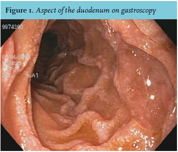
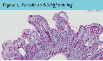
A colonoscopy, gastroscopy, and an abdominal CT were then performed. No abnormalities were seen on colonoscopy. Gastroscopy showed a remarkable erosive feature of the duodenum (figure 1), which was biopsied (figure 2). Abdominal CT showed retroperitoneal and particularly mesenteric lymphadenopathy, and thickened intestinal walls in the proximal jejunum.
WHAT IS YOUR DIAGNOSIS?