Full textPDF
Full text
CASE REPORT
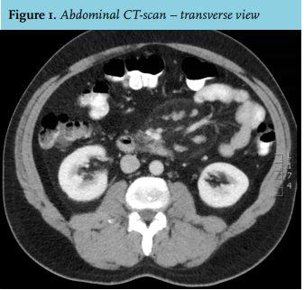
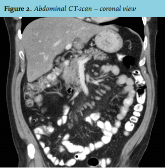
A 43-year-old man presented to our emergency department for the second time in three days because of abdominal pain, vomiting and lack of defecation. His medical history revealed a sliding hiatal hernia and reflux oesophagitis for which he took pantoprazole 40 mg twice daily. The abdominal pain started in the left lower quadrant four days before the second presentation, but had now spread through the entire abdomen with maximal intensity in the right lower quadrant. Paracetamol and laxatives did not reduce the pain sufficiently. Physical examination did not reveal any abnormalities and he was afebrile. Laboratory tests including liver enzymes, renal function and inflammation parameters were within the normal ranges. Abdominal X-ray showed a normal bowel gas pattern and faecal material. Additionally, an abdominal computed tomography scan was performed (figures 1 and 2).
WHAT IS YOUR DIAGNOSIS?