INTRODUCTION
Well-known side effects of doxycycline are photosensitivity, teeth discolouration, nausea, vomiting, and diarrhoea. Cutaneous hyperpigmentation is a common side effect of minocycline and, to a lesser extent, of other tetracyclines, with only one report of a patient with progressive, symmetric blue-grey periocular discolouration due to three years of treatment with therapeutic doses of doxycycline.1 Furthermore, hyperpigmentation has been described in one patient with self-induced intoxication by doxycycline (1 gm/day) for 12 years.2 Both brown discolouration of the fingernails and discolouration of acne scars have been described after a short course of doxycycline.3,4 We report four patients who received long-term treatment with doxycycline and hydroxychloroquine because of either chronic Q-fever or Whipple’s disease. They showed extensive cutaneous hyperpigmentation in previously unaffected skin, probably induced by doxycycline.
|
What was known on this topic? What does this add? |
|
|
CASE DESCRIPTIONS
Case 1
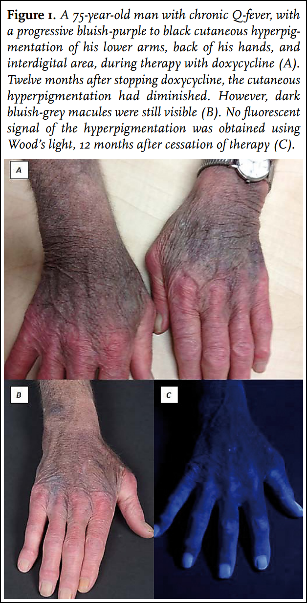
A 75-year-old man with an abdominal aneurysm, immunosuppressive therapy because of rheumatoid arthritis and a known valvulopathy was diagnosed with chronic Q-fever. Doxycycline 200 mg/day was initiated, in addition to hydroxychloroquine 400 mg/day, which he had already been taking for more than five years because of rheumatoid arthritis. After four months, doxycycline 300 mg/day was introduced because of persistently low doxycycline levels. Eight months after the start of therapy, progressive bluish-purple to black cutaneous hyperpigmentation of his lower arms, back of his hands, and interdigital areas (figure 1A) developed since increasing the doxycycline dose (serum concentrations of 5.8 mg/ml). The doxycycline was stopped and hydroxychloroquine was continued. The hyperpigmentation slowly diminished, but 12 months later dark bluish-grey macules were still visible on the back of his hands and his lower arms (figure 1B).
Case 2
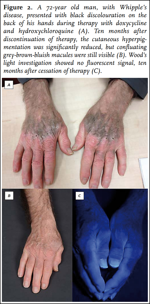
A 72-year-old man, diagnosed with relapse of Whipple’s disease, was treated with ceftriaxone for four weeks, followed by doxycycline 200 mg/day and hydroxychloroquine 600 mg/day. Eight months later, increasing black discolouration on the back of both hands was seen (doxycycline serum concentrations of 5.7 mg/ml) (figure 2A). Therapy was stopped, and co-trimoxazole was reintroduced. Ten months later his cutaneous hyperpigmentation was slowly fading (figure 2B).
Case 3
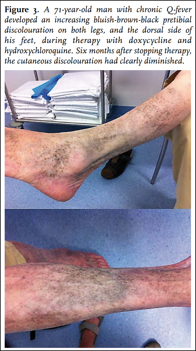
A 71-year-old man with an endovascular aneurysm repair (EVAR) and a femoral-popliteal bypass was referred because of aortitis due to chronic Q-fever, and started on doxycycline 200 mg/day and hydroxychloroquine 600 mg/ day. After 48 months of therapy, he reported increasing pretibial bluish-brown-black discolouration on both legs, and the dorsal side of his feet (figure 3). In retrospect, the discolouration started 11 months before, but he had never reported it. Doxycycline and hydroxychloroquine were substituted by moxifloxacin and rifampicin. Six months later, the discolouration diminished.
Case 4
A 72-year-old man with an infected EVAR with retroperitoneal abscesses due to chronic Q-fever was referred for surgery. He had already received six months of doxycycline 300 mg/day and hydroxychloroquine 600 mg/ day (doxycycline serum concentration: 6.2 mg/ml), which was continued post-surgery. For six months, he received doxycycline 200 mg/day because of side effects. However, because of a low doxycycline serum concentration (2.8 mg/ ml), doxycycline 300 mg/day was reintroduced, leading to a near-therapeutic concentration (4.7 mg/ml). Eight months post-surgery, he presented with increasing black discolouration around the surgical scars on both legs. Doxycycline and hydroxychloroquine were substituted by moxifloxacin. Two months later, the black discolouration diminished.
DISCUSSION
We describe four patients with hyperpigmentation of previously healthy skin after prolonged use of doxycycline. This has been described before in only one patient with therapeutic doses of doxycycline,1 and in a patient with self-induced doxycycline intoxication (1 g/day during 12 years leading to doxycycline serum concentrations of 34 mg/ml, normal therapeutic range: 1-5 mg/ml, for chronic Q-fever: 5-10 mg/ml).2,5 In our cases, patients received relatively high doses with serum concentrations in the therapeutic range, and developed marked cutaneous hyperpigmentation. However, compared with other indications for which doxycycline is given, chronic Q-fever and Whipple’s disease require prolonged treatment with a higher therapeutic range. Because tetracyclines produce autofluorescence, with positive in-vivo conjunctival autofluorescence of palpebral conjunctival minocycline deposits,6 the hyperpigmentation of the first two cases was investigated with Wood’s light (extinction 365 nm). However, no fluorescent signal was obtained (figures 1C and 2C). This may have been due to the long time that elapsed between the cessation of doxycycline and this investigation (12 and 10 months, respectively). As the dorsal side of the hands of the first patient still showed clear pigmentations (figure 1B), the pigment might not represent the doxycycline itself. Previously, biopsies of doxycycline-induced hyperpigmentation revealed increased melanisation in the basal layers of the epidermal keratinocytes,4,5 suggesting activation of melanocytes either by the tetracycline derivative itself or by another co-stimulus. Also, indications were found for the presence of melanin or melanin-like pigment in the histiocytes of the upper dermis. In contrast, in histiocytes of the lower dermis and subcutaneous fat, pigment was stored with increased amounts of iron and calcium, and no melanosomes were detected, suggesting a different nature of the pigment. Furthermore, data suggested that doxycycline, possibly chelated with iron and/or calcium, was directly deposited in the lesional skin.5 The role of hydroxychloroquine and its interaction with doxycycline in these cases cannot be completely ruled out, as cutaneous hyperpigmentation induced by hydroxychloroquine has been described in 13% of treated patients, mainly as a bluish-grey pigmentation,7 mostly localised at the hard palate, gums, face, and pretibial area.8 To our knowledge, no literature exists describing an increased risk of hyperpigmentation using doxycycline and hydroxychloroquine concomitantly. As both medications can cause cutaneous hyperpigmentation a synergistic effect on the development of hyperpigmentation might exist. However, based on the localisation of hyperpigmentation, without mucosal involvement,9,11 doxycycline is still thought to be the main aetiological agent in our cases. Furthermore, in the first patient, hyperpigmentation developed after introduction of doxycycline 300 mg/day, and significantly diminished after stopping doxycycline, while hydroxychloroquine was continued. And, as seen in our fourth patient, discolouration restricted to scars has been reported with doxycycline.4 Most described cases of cutaneous hyperpigmentation during tetracycline treatment are induced by minocycline,12 which is frequently prescribed for long periods. However, indications for prolonged therapy with doxycycline also exist, with an increasing number of chronic Q-fever patients.13 It should be advised to discontinue therapy. As in our patients, partial to complete resolution of cutaneous hyperpigmentation has been described eight months after cessation of prolonged doxycycline therapy.1 Furthermore, in the case with doxycycline intoxication, the pretibial hyperpigmentation had faded significantly one year after doxycycline cessation.2 Finally, almost complete disappearance of methacycline-induced hyperpigmentation was reported five years after onset, except for two patients who were substituted with doxycycline.14 Complete disappearance of hyperpigmentation after cessation of therapy is possible; however, recovery may take up to several years.14
In conclusion, cutaneous hyperpigmentation is a potential side effect of doxycycline therapy within the therapeutic dose range, and the chance to evoke this adverse effect might be increased with the concomitant use of hydroxychloroquine. Given the widespread use of doxycycline, in both short and prolonged regimens, it is important to recognise this reversible or partially reversible side effect in order to discontinue therapy. Especially its use in chronic Q-fever, when prolonged relatively high doses are given nowadays in combination with hydroxychloroquine, prescribers and patients should be aware of this side effect.
DISCLOSURES
The authors declare that they have no competing interests. The authors declare that the final manuscript has not been submitted or accepted for publication elsewhere. This work received no financial support.
A C K N O W L E D G E M E N T S
The authors would like to thank A. Prischmann, medical photographer from the department of Dermatology, Radboudumc in Nijmegen, the Netherlands, for her contribution in image formation of the affected skin.
REFERENCES
1. Pichardo RO, Yeatts RP, Sangueza OP. Doxycycline-inducted Cutaneous Hyperpigmentation (Abstract only). Am J Dermatopathol. 2006;28:235.
2. Westermann GW, Bohm M, Bonsmann G, Rahn KH, Kisters K. Chronic intoxication by doxycycline use for more than 12 years. J Intern Med. 1999;246:591-2.
3. Akcam M, Artan R, Akcam FZ, Yilmaz A. Nail discoloration induced by doxycycline. Pediatr Infect Dis J. 2005;24:845-6.
4. Adisen E, Gurer MA, Erdem O. Tetracycline/doxycycline-induced cutaneous depressed pigmentation. Int J Dermatol. 2006;45:1245-7.
5. Bohm M, Schmidt PF, Lodding B, et al. Cutaneous hyperpigmentation induced by doxycycline: histochemical and ultrastructural examination, laser microprobe mass analysis, and cathodoluminescence. Am J Dermatopathol. 2002;24:345-50.
6. Lim LT, Tarafdar S, Collins CE, Roberts F, Ramaesh K. Minocycline induced conjunctival autofluorescence deposition. Semin Ophthalmol. 2012;27:25-6.
7. Reynaert S, Setterfield J, Black MM. Hydroxychloroquine-induced pigmentation in two patients with systemic lupus erythematosus. JEADV. 2006;20:487-8.
8. Skare T, Ribeiro CF, Souza FH, Haendchen L, Jordao JM. Antimalarial cutaneous side effects: a study in 209 users. Cutan Ocul Toxicol. 2011;30:45-9.
9. Ochsendorf FR, Runne U. [Chloroquine and hydroxychloroquine: side effect profile of important therapeutic drugs]. Hautarzt. 1991;42:140-6. Chloroquin und Hydroxychloroquin: Nebenwirkungsprofil wichtiger Therapeutika.
10. Fardet L, Revuz J. [Synthetic antimalarials]. Ann Dermatol Venereol. 2005;132:665-74. Antipaludeens de synthese.
11. Koranda FC. Antimalarials. J Am Acad Dermatol. 1981;4:650-5.
12. Klein NC, Cunha BA. Tetracyclines. Med Clin North Am. 1995;79:789-801.
13. van der Hoek W, Schneeberger PM, Oomen T, et al. Shifting priorities in the aftermath of a Q fever epidemic in 2007 to 2009 in The Netherlands: from acute to chronic infection. Euro Surveill. 2012;17:20059.
14. Moller H, Rausing A. Methacycline hyperpigmentation: a five-year follow-up. Acta Derm Venereol. 1980;60:495-501.